We use our muscles for practically everything, and problems with the muscular system can affect us in a variety of different ways. These issues can range from mild discomfort to muscle loss and in extreme cases death. Located under the term Musculoskeletal Diseases (MSD), muscle-related diseases are a major contributor to disability worldwide, with their prevalence rising even further with aging populations.
To date, new therapies and drugs for MSD have been limited by a lack of 3D functional and reliable in vitro tissue models, with more widely used 2D cell culture systems suffering from inherent flaws. Typically, grown in a monolayer on a flat surface, 2D cultured cells cannot truly reflect the way human tissue structures grow or respond in the human body.
Running alongside 2D cell cultures, another tool often used in research involves the testing of drugs on animal models, which raises a variety of ethical and economical concerns. Furthermore, the translation of results obtained from animal experiments to humans is problematic. And despite being capable of overcoming the shortcomings of both 2D and animal models, past 3D cell culture processes have been labeled as being too time-consuming, cumbersome and difficult to replicate or scale-up.
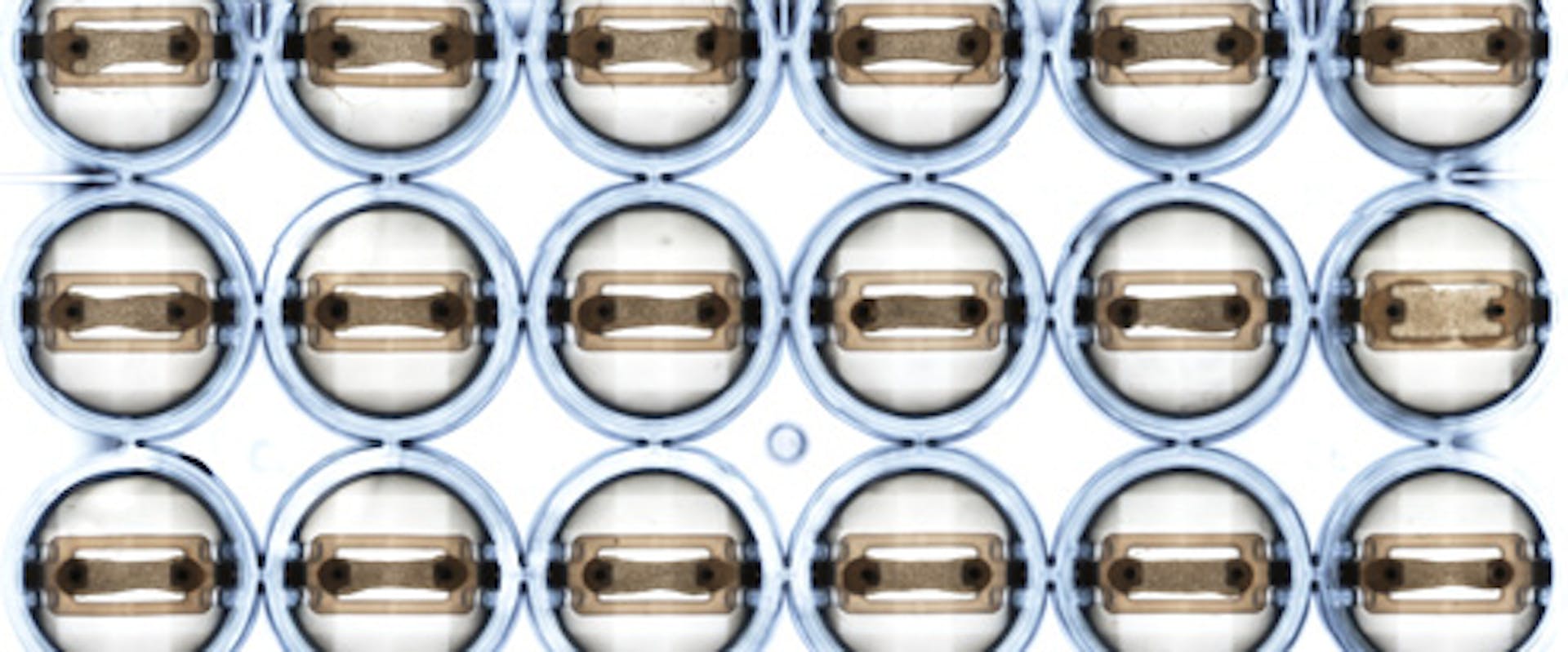
Compact in size, ‘MUSTANG’ can analyze 3D bioprinted human muscle tissues within a 24-well plate. After undergoing maturation and differentiation in an incubation chamber, electrodes stimulate the micro-muscles, mimicking the biological functionality of the tissues (e.g. force and fatigue). All the while, the differences in the way these tissues contract and react in the presence of different drugs or compounds such as caffeine (a well-known muscle stimulant) are automatically and optically monitored by an advanced algorithm developed at CSEM.
“The forces we measure via the algorithm are absolutely minute, I guess they could be compared to measuring the weight of a house fly in the order of 100 - 500 μN,” describes Vincent Revol. CSEM was challenged with the development of “The force readout module – solving the challenge of keeping the muscles in their incubation environment while measuring the force exerted. For this, we designed an optical module and used a position tracking algorithm based on machine learning – allowing us to measure positions robustly at sub-micrometer resolution.”
“Thanks to the precise and accurate nature of this data collection, the 3D tissue analyzer platform will be able to test new drugs for MSD more efficiently, hopefully leading to novel therapies for patients,” says Hansjoerg Keller, Senior Principal Scientist at Novartis. “In the not too distant future we would like to extend the platform to cardiac muscles enabling us to address an even broader range of pharmaceutical applications,” he adds.