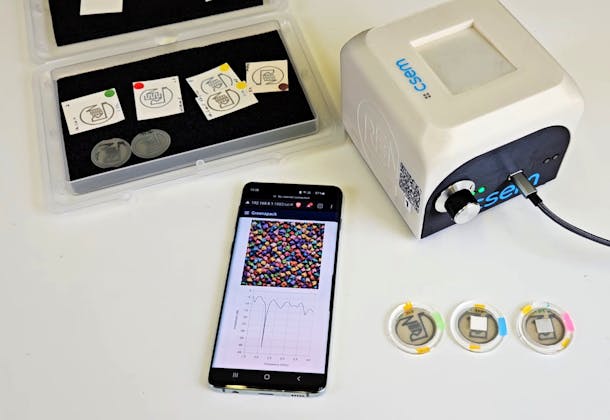
Scanning electron microscope picture of an array of nano cones. Each nano cone is located inside a nano cavity. Electrons are extracted from the nano cones by applying a voltage to the top gate electrode.
Microfabrication with Swiss precision
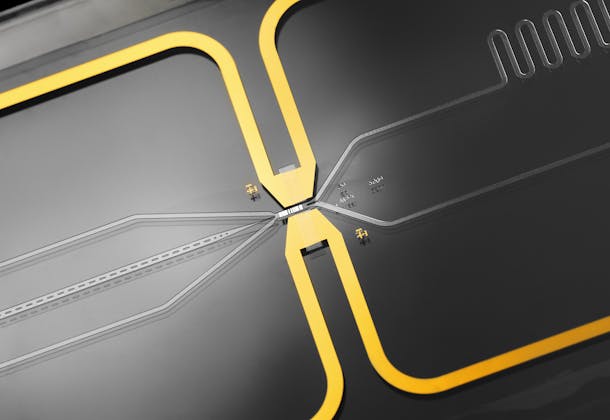
CSEM, in partnership with Nanox, has now been capable of producing fully functional MEMS chips to be integrated into the Nanox.ARC system and other applications as well. The MEMS chip consists of millions of homogeneous nano-cones, regularly spaced and uniformly shaped refractory metal cones – each only a few hundred nanometers in size – arranged on an area of about seven square millimeters. For electron emission, a positive voltage is applied to the grid and pulls a stream of electrons from the tips of the cones.
“For the generation of millions of nano-cones of exactly the same height we had to develop a unique electro-chemical etching,” says senior project leader Thomas Overstolz, Senior R&D Engineer at CSEM. “The etching process had to be very precise and timed to the second.” The production took place at the state-of-the-art cleanroom of the specialized MEMS foundry at CSEM.
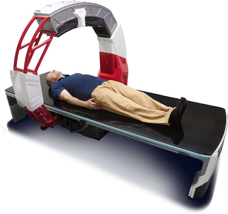
Nanox.ARC: 3D tomosynthesis imaging system.
MEMS chip facilitates breakthrough medical imaging
The innovative Nanox.ARC system developed by NANOX has several advantages compared to the conventional imaging system. As mentioned, a tungsten filament must be heated to 2000°C. In contrast, the cold cathode tube does not need a standby current and can be turned on and off instantly with low voltage and milliamps current consumption. Also, because there is no need for a standby current, several vacuum tubes (or even several chips within a single tube) can be placed closely together while connected to the High kV and switched one by one with no X-ray radiation from the neighboring source. This is not possible to achieve with hot-cathode sources. Nanox intends to build tubes with CSEM chips in them, for testing and validation in the next stage.
The novel digital X-ray source is the basis of core technology in the imaging systems Nanox is developing. Nanox’s mission is to improve early detection and treatment, which they believe is key to helping people achieve better health outcomes Their goal is to enable medical institutions to incorporateend-to-end systems, or to adopt a modular approach by acquiring or licensing components to integrate Nanoxtechnologies into their specific products.